SOLUTIONS FOR MUSCLE TISSUE ASSAYS
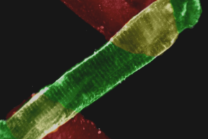
Lying between single cell and whole heart experimental studies, intact muscle tissue isolated from hearts offers unique advantages. Unlike single isolated myocytes, which represent the smallest fully functional model system, intact muscle can be used to study cardiac function while preserving the native myocardial tissue’s multicellular environment, including both fibroblasts and myocytes. And unlike whole heart studies, the contractile characteristics of muscle tissue can be evaluated independently of extrinsic factors such as vascular tone and are better suited for acquisition of functional parameters like calcium transients. Muscle tissue preparations also allow measurements too difficult or impossible to perform in whole hearts. Similarly, unlike skinned preparations, intact muscle preparations allow for simultaneous detection of force production and intracellular calcium dynamics.
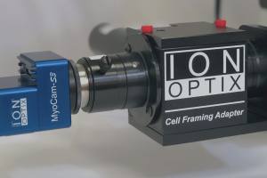
We offer two variations of the same core system featuring quantitative force and fluorescence measurements, as well as real-time force-length work loop data acquisition. Our MyoClamp System permits either electrical excitation directly through the platinum hooks attached to the muscle preparation as well as electrical field stimulation. The system’s flexible design allows it to accommodate a variety of tissue types including primary and engineered, as well as cardiac and skeletal tissues. And our systems also include many of the key tools required for successful tissue preparation.


IonOptix offers turn-key solution for studying intact muscle tissue…

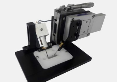
MYOCLAMP SYSTEM
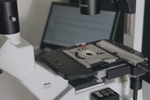
The IonOptix MyoClamp System enables measurements of calcium, force, and mechanical work in thin slice preparations of myocardium, intact ventricular papillary muscle, and other excitable primary and engineered muscle tissue. Thin slice preparations are adhered to stable clips, then mounted between a robust force transducer and programmable length controller in a perfusion chamber, allowing for continuous temperature control and oxygenation of the tissue. The chamber also includes fixed, inert platinum electrodes providing electrical field stimulation while minimizing electrolysis. Other tissue types are mounted via platinum omega clips; our specialized force transducer permits electrical excitation directly through the platinum hooks attached to the muscle preparation, ensuring no electrolysis. LEARN MORE