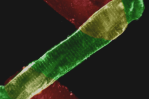
Lying between single cell and whole heart experimental studies, cardiac slices provide many of the benefits of a reductionist approach of isolated cardiac myocytes while being easier to generate and more robust with added physiological relevance.
Cardiac slices, unlike single isolated myocytes, can be used to study cardiac function within a multicellular context and an intact myofilament lattice. Slices have the added benefit of maintaining a syncytium of myocytes found in vivo thus maintaining in vivo architecture and intercellular signaling, suggesting that experimental results are more likely to have physiological relevance. And unlike whole heart studies, cardiac slices allow measurements too difficult or impossible to perform in whole hearts.
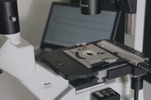
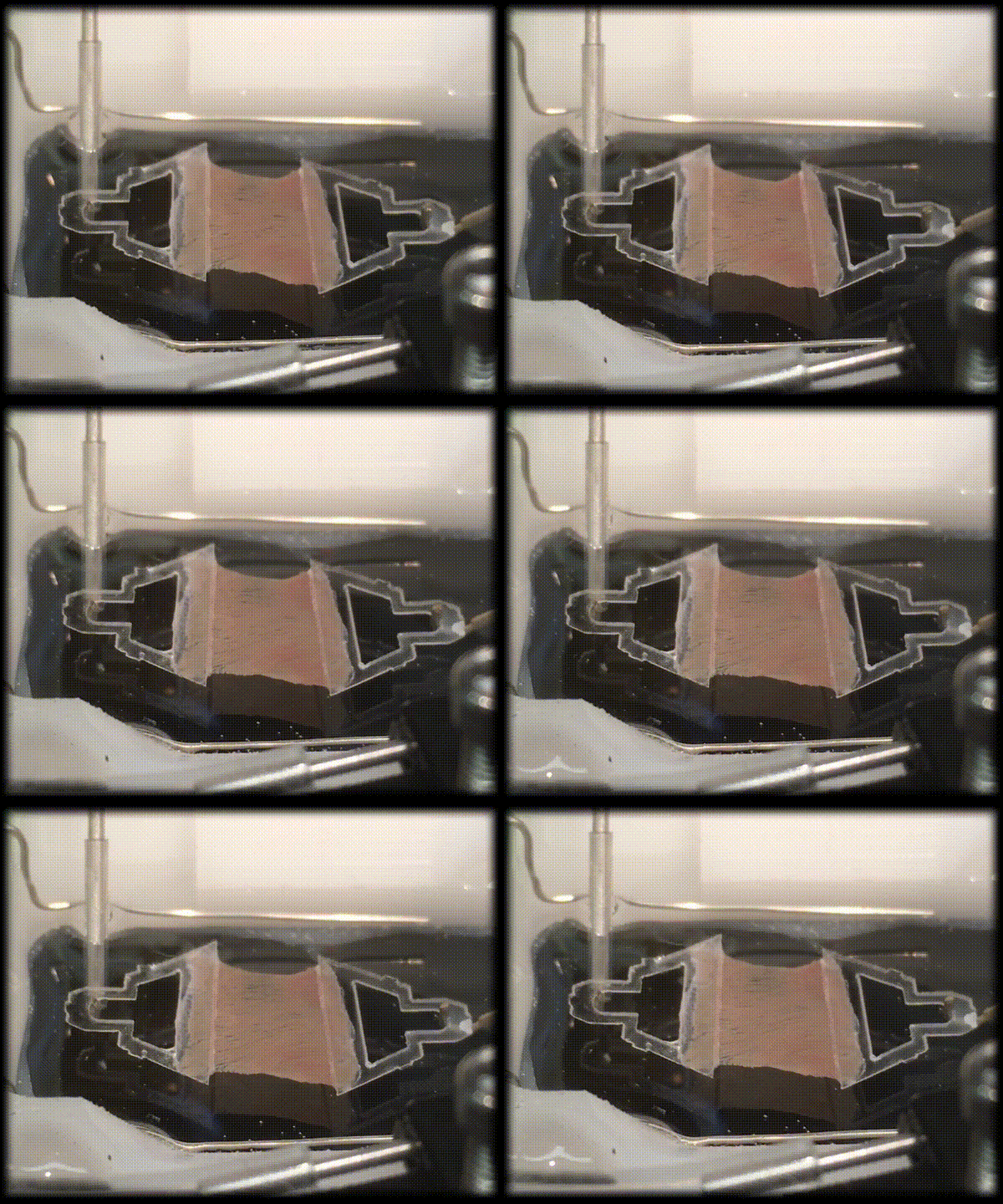
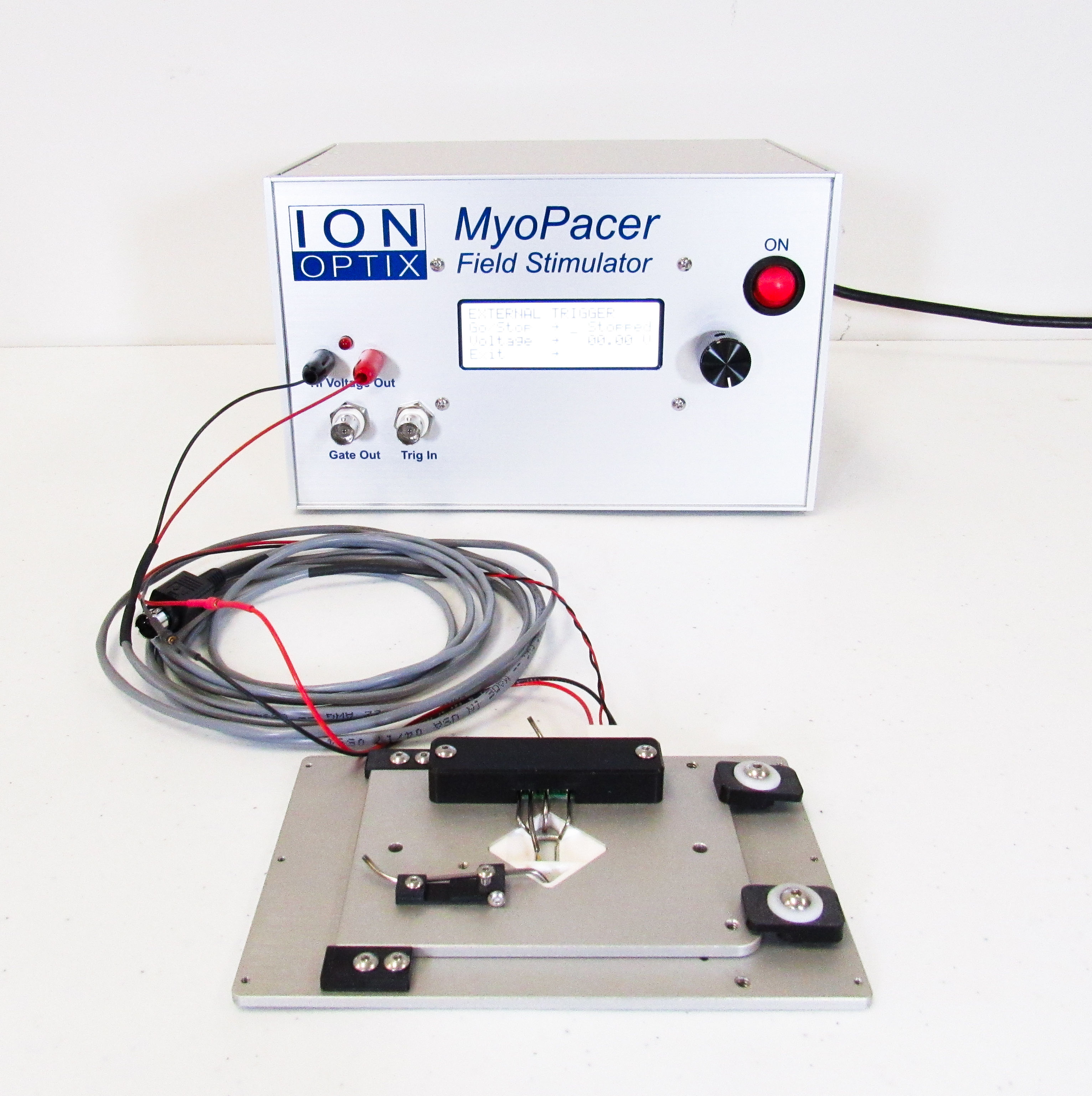
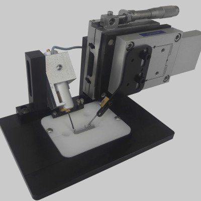
The IonOptix Cardiac Slice System has been designed to facilitate the application from slice preparation to data acquisition and analysis. Cardiac slices are easily affixed to sturdy, stable triangular clips that mount between a robust force transducer and programmable length controller. Chamber fluid flow allows for temperature control and continuous oxygenation of tissue, while fixed platinum electrodes enable electrical field stimulation. The platinum allows for electrical conductance and are biologically inert while minimizing electrolysis.
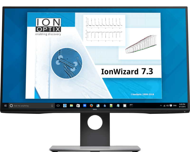
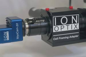
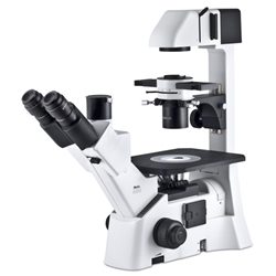
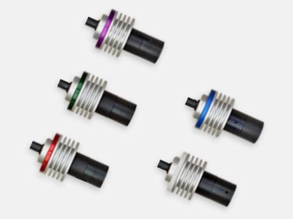
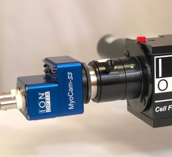
When combined with the IonOptix Calcium and Contractility System, fluorescence of calcium-sensitive dyes, such as Fluo-4, Fura-2 and/or Indo-1, can be detected using IonWizard, which permits control of experiment acquisition parameters and analysis of data.
Similar to pressure-volume work loops in whole heart, mechanical work loops generated by cardiac slices (displayed in real-time) can be acquired using force-feedback length control. Force-length work loops can be used to derive important functional data including end-systolic and end-diastolic force-length relationships (ESFLR and EDFLR) characterizing the elastance and compliance of the tissue and mechanical work and power.
The cardiac slice preparation technique has been recently championed by Professor Cesare Terracciano (Imperial College London) and his laboratory, including Dr. Fotios Pitoulis. We would like to acknowledge and thank them for their assistance with the tissue preparation.
As with all of our systems, we stand behind our products. When we install our complete systems we use your preparations to help get you started as quickly as possible. And when you need assistance we offer unlimited phone and email support for the lifetime of your system.
SPECIFICATIONS
FEATURES
COMPONENTS
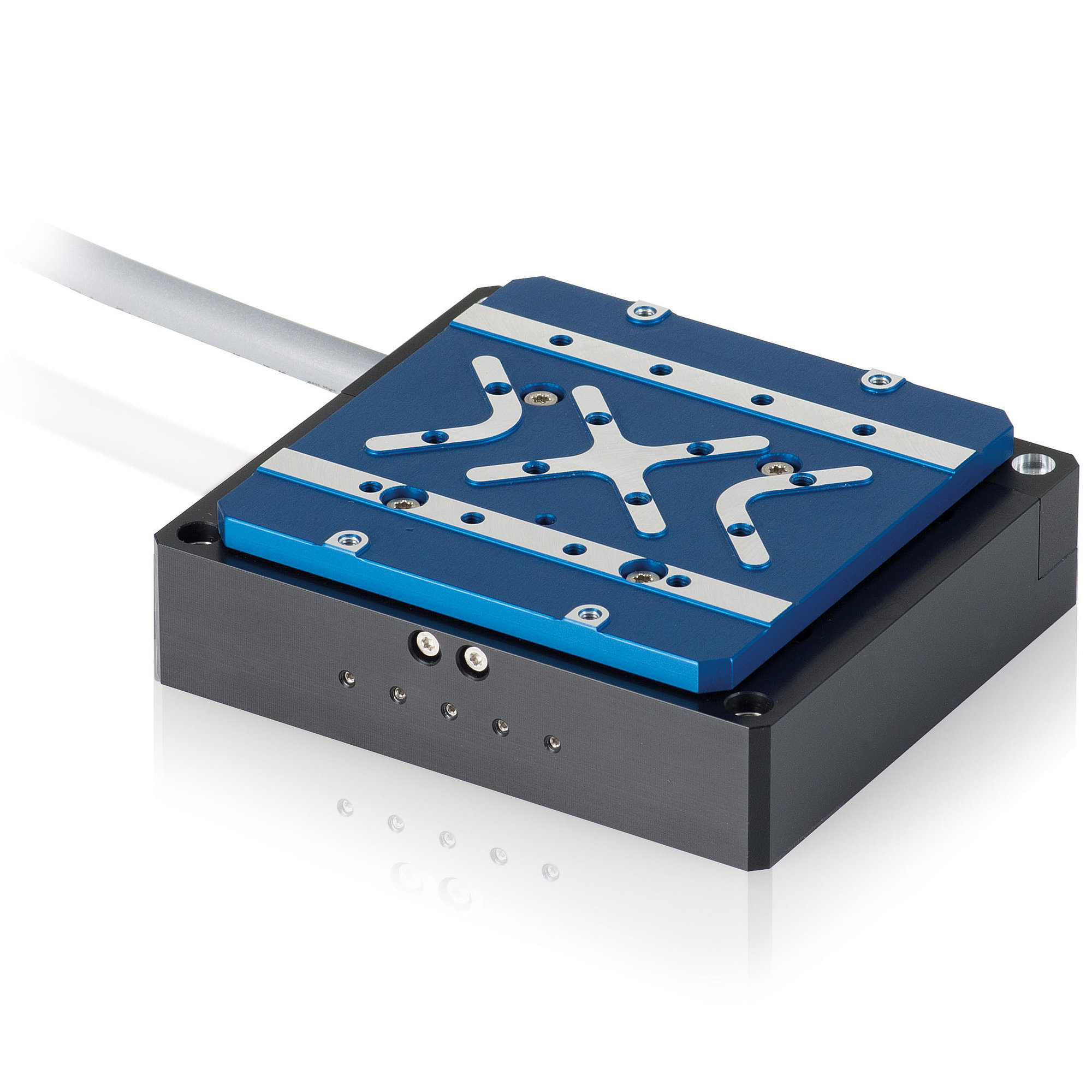
Cardiac Slice Systems can be combined with our typical Calcium and Contractility components to give a full suite of simultaneous fluorescence and contractility (i.e. force development) measurements. Depending on your specific application, systems may include a sensitive force transducer and/or a programmable length controller.
BROWSE THE TABS to learn more about the components that complete this system.
SOFTWARE
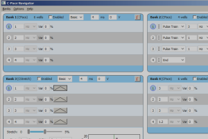
Data acquisition in the muscle system is achieved through the core IonWizard software, in which trace recordings display force data and length controller position. Add the software module PMTAcq for fluorescence recording, the Advanced Signal Generator for precise muscle length control with programmable protocols, and/or MultiClamp for force-length work loops.
